Overview
The life history of an individual coral is archived within the accreting skeleton of the colony. While reef-forming coral colonies (e.g. massive Porites sp.) may live for hundreds of years and deposit calcareous structures many metres in height and width, their living tissue is a thin outer surface layer comprised of asexually-dividing polyps that only survive a few years. To understand the rate and timing of polyp division and the consequences for colony skeletal growth, scientists need to track the skeletal corallite deposited around each polyp.
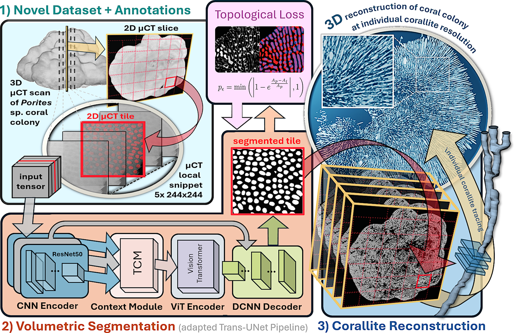
CoralLite is an annotated μCT scan dataset of entire calcareous skeletons and an associated, first corallite deep learning reconstruction baseline. CoralLite combines fully quantified volumetric segmentations with cross-slice linking for visualisations of 3D models for each corallite up to colony scale. For segmentation, we propose and evaluate a hybrid V-Trans-UNet architecture applicable to segmenting tiled μCT virtual slabs of Porites sp. colonies. The model is pre-trained on weakly annotated data and topology-aware fine-tuned using fully annotated slice sections with 8k+ manual corallite region annotations.
On unseen slices of the same colony, the resulting model reaches 0.94 topological accuracy at mean Dice scores of 0.77 on the same colony and projection axis, and 0.63 mean Dice on a different, biologically unrelated specimen. Our results show for the first time that visual machine learning can effectively support full 3D individual corallite modelling from μCT scans of coral skeletons alone.
We publish the full dataset of 697 μCT slices, 37 partial or full slice annotations, and all network weights and source code with this work to enable reproducibility and future research.
Download: Zenodo — 10.5281/zenodo.21935696 (DOI reserved; resolves on publication)
Dataset
CoralLite comprises high-resolution μCT data from three Porites specimens from the Natural History Museum (London, UK) and Naturalis Biodiversity Center (Leiden, NL), scanned using a Nikon XT H 225 ST system.
| Specimen | Slices | Annotation | Usage |
|---|---|---|---|
| Naturalis 6785 | 60 | 33 partial (centre points) | Pre-training |
| Naturalis 6781 Growth | 248 | Full slices (#1279, #2499) | Fine-tuning / test |
| Naturalis 6781 Ortho | 388 | Full slice (#1003) | Cross-axis test |
| Astraeiformis (NHM) | 1 | Full slice | Cross-species test |
In total the release includes 8,412 manually annotated corallite interior regions across four fully annotated slices, complemented by weak point annotations on 33 additional slices. Six annotators produced weak labels under expert oversight; four slices provide pixel-level binary masks for training and evaluation.
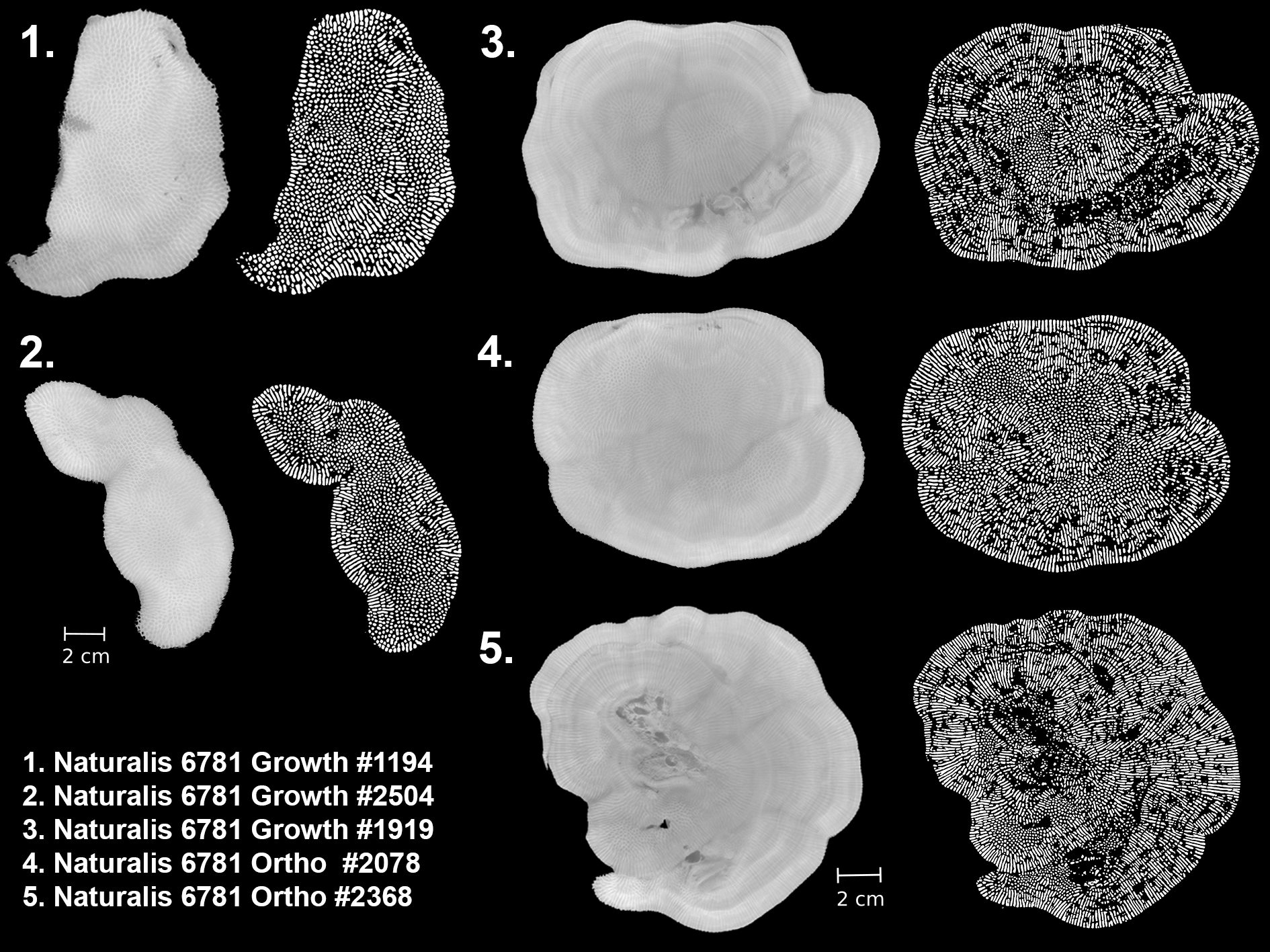
Example colony slices and corresponding segmentation outputs across growth and orthogonal projection axes (Naturalis 6781).
Evaluation tasks
- Task A: same colony, same projection axis (Naturalis 6781 Growth #2499)
- Task B: same colony, different axis (Naturalis 6781 Ortho #1003)
- Task C: entirely different specimen (Porites astraeiformis, NHM)
Methods
Volumetric segmentation pipeline
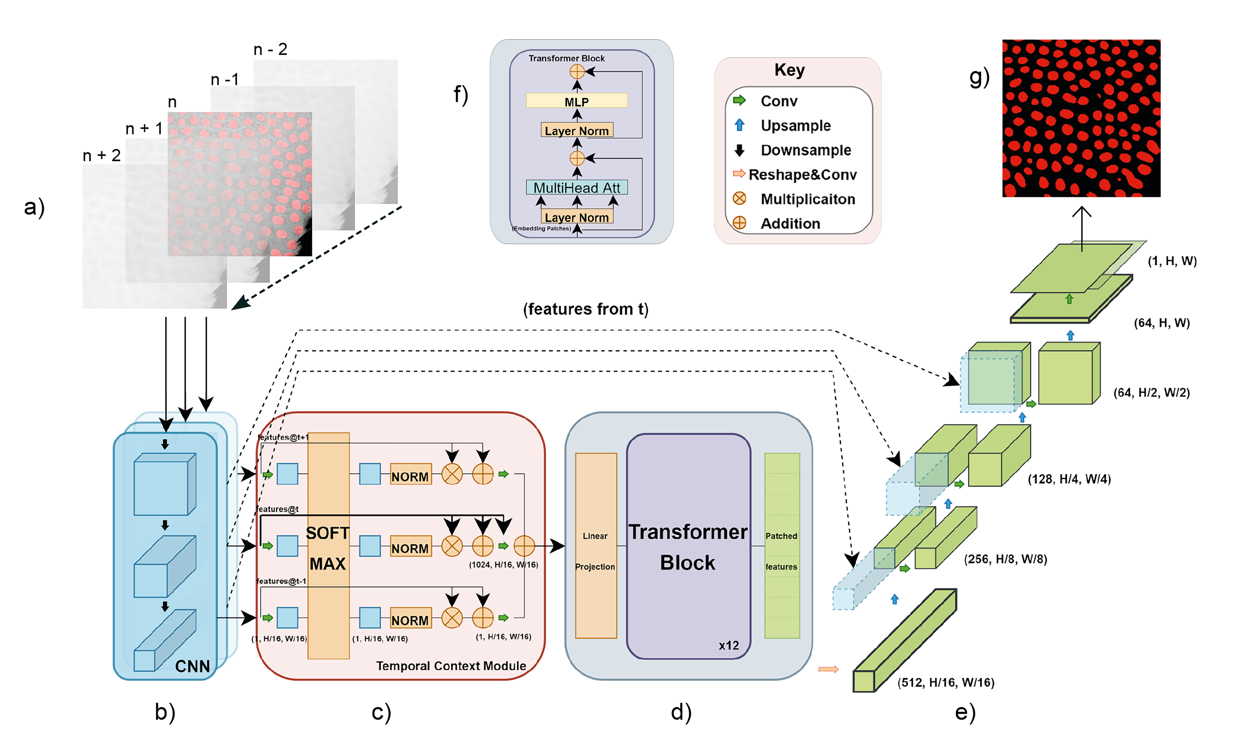
Each μCT slice is processed using overlapping 224×224 tiles. Training samples are 5×224×224 volumetric snippets (centre-slice supervision) to exploit continuity across adjacent slices. We adapt Video-Trans-UNet: a ResNet-50 encoder, temporal context module, 12-layer ViT, and UNet decoder with skip connections.
Topology-aware loss
Standard Dice/BCE losses do not penalise merged neighbouring corallites — a critical failure mode for downstream 3D tracing. We add a connectivity-aware penalty via connected-component matching, applied during fine-tuning with coefficient T = 0.1 after warm-up. Training is two-stage: weak-label pre-training on Naturalis 6785, then fine-tuning on a single fully annotated slice from the target colony.
3D corallite reconstruction
Predicted 2D regions are linked across slices using centroid distance and intersection-over-union matching to form identity-preserving 3D corallite tubes suitable for colony-scale visualisation.
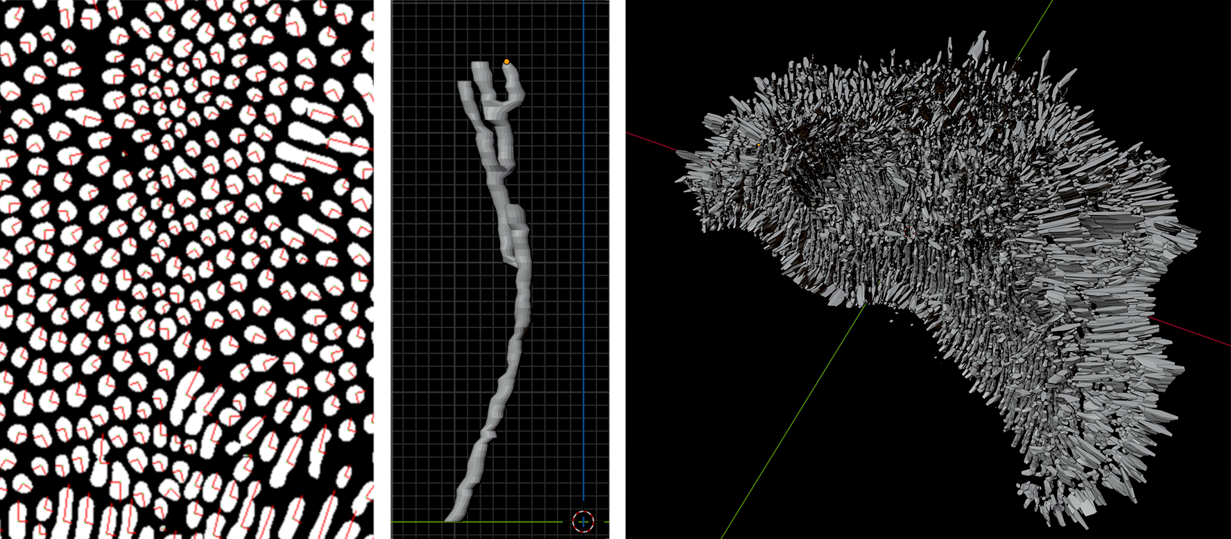
Per-slice detection, individual corallite tracing, and colony-scale 3D visualisation.
Results
Fine-tuning on one annotated slice is sufficient for strong same-colony performance. The best configuration combines dense tiling (k = 50), topology loss (T = 0.1), and Gaussian noise augmentation:
| Model | Task A (mDSC / Topo) | Task B (mDSC / Topo) | Task C (mDSC / Topo) |
|---|---|---|---|
| Pre-training only | 0.01 / 0.66 | 0.01 / 0.75 | 0.01 / 0.68 |
| Fine-tuned (k=50) | 0.76 / 0.94 | 0.71 / 0.93 | 0.56 / 0.79 |
| + Topo + noise (best) | 0.77 / 0.94 | 0.71 / 0.93 | 0.63 / 0.87 |
Cross-species generalisation remains the hardest setting; topology loss improves structural separation and noise augmentation reduces scanner-echo false positives.
Citation
@inproceedings{jones2026corallite,
  title     = {{CoralLite}: $\mu$CT Reconstruction of Coral Colonies from Individual Corallites},
  author    = {Jones, Jess and Bertini, Leonardo and Johnson, Kenneth and Hendy, Erica and Burghardt, Tilo},
  booktitle = {Proceedings of the CVBMC Workshop at the International Conference on Pattern Recognition (ICPR)},
  year      = {2026},
  url       = {https://icpr2026orgteam.github.io/WSpapers/pdfs/paper_0164.pdf}
}

@dataset{jones2026corallite_data,
  author    = {Jones, Jess and Bertini, Leonardo and Johnson, Kenneth and Hendy, Erica and Burghardt, Tilo},
  title     = {{CoralLite}: $\mu$CT Coral Dataset and Model Weights},
  year      = {2026},
  publisher = {Zenodo},
  doi       = {10.5281/zenodo.21935696}
}
Acknowledgments
J.J. is supported by the FARSCOPE EPSRC CDT. J.J. and T.B. acknowledge the University of Bristol HPC facilities. E.H. and L.B. thank the dedicated labelling of corallites completed by University of Bristol Environmental Geoscience undergraduates (Arisha Fancy, Poppy Hammond, Sharifah Nor Qistina Syed Izuan, Louisa Lou, Emma Mody, and Khalil Imran Abdul Rahman), funded through the NERC GW4 DTP Undergraduate Research Experience Placement scheme and the University of Bristol School of Earth Sciences Summer Internship programme. Support for L.B. is acknowledged through 4D-REEF (Marie Skłodowska-Curie ITN, grant 813360). We are grateful for support from SYNTHESIS+ Transnational Access project NL-TAF-TA3-009, the Natural History Museum Science Investment Fund, and the University of Bristol Covid Recovery Fund.
Specimens were provided by the Natural History Museum, London and Naturalis Biodiversity Center, Leiden.
Contact: jess.jones@bristol.ac.uk · tilo@cs.bris.ac.uk